Structure and Function
The sense of smell (olfaction) monitors chemicals and determine the flavor and palatability of foods and beverages.
It warns of dangerous environmental conditions, including fire, air pollution, leaking natural gas, and bacteria-laden foodstuffs.
Olfactory pathway, and the connection of the pathway to memory (and behavior) related brain regions.
-
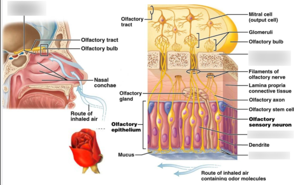
Olfactory Epithelium
-
Located in the roof of the nasal cavity, superior nasal concha, and upper part of the nasal septum.
-
Specialized pseudostratified epithelium containing:
-
Olfactory receptor neurons (ORNs): Bipolar neurons with cilia that detect odorants.
-
Supporting (sustentacular) cells: Provide structural and metabolic support.
-
Basal cells: Stem cells that regenerate olfactory neurons (unique among neurons).
-
Bowman’s glands: Secrete mucus to dissolve odorants.
-
-
- Olfactory Receptor Neurons
Bipolar neurons with dendrites extending into the mucus layer.
-
Cilia on the dendrites contain odorant receptor proteins.
-
Axons of ORNs (unmyelinated) bundle together as olfactory nerve filaments (CN I) and pass through the cribriform plate of the ethmoid bone to reach the olfactory bulb.
-
- Olfactory Bulb
-
First relay station for olfactory information, located on the ventral surface of the frontal lobe.
-
Contains structures called glomeruli, where axons of receptor neurons synapse with dendrites of mitral and tufted cells (secondary neurons).
-
Also contains periglomerular and granule cells, which provide inhibitory modulation.
-
- Olfactory Tract
-
Formed by axons of mitral and tufted cells.
-
Projects to the primary olfactory cortex and related limbic structures.
-
Divides into medial and lateral striae:
-
Lateral stria: Major pathway to olfactory cortex (piriform cortex, amygdala, entorhinal cortex).
-
Medial stria: Projects to contralateral olfactory bulb via anterior commissure.
-
-
- Central Olfactory Pathways
-
Primary olfactory cortex: Piriform cortex, periamygdaloid cortex, and entorhinal cortex (temporal lobe).
-
Secondary olfactory areas: Orbitofrontal cortex (involved in odor discrimination and conscious perception).
-
Strong connections with the limbic system (amygdala, hippocampus) explain why odors are closely linked to memory and emotions.
-
Physiology of the Olfactory System
-
Odor Detection
-
Odorants are inhaled and dissolve in the mucus layer of the olfactory epithelium.
-
They bind to G-protein–coupled receptors (GPCRs) on the cilia of receptor neurons.
-
Each receptor neuron expresses only one type of receptor, but each receptor can bind multiple odorants.
-
-
Signal Transduction
-
Odorant binding activates a G-protein (Golf).
-
This stimulates adenylate cyclase, increasing cAMP levels.
-
cAMP opens cyclic nucleotide-gated (CNG) ion channels, allowing Na⁺ and Ca²⁺ influx.
-
Ca²⁺ opens Cl⁻ channels, amplifying depolarization.
-
If threshold is reached, an action potential is generated in the olfactory receptor neuron.
-
-
Neural Processing
-
Axons of ORNs converge onto glomeruli in the olfactory bulb.
-
Each glomerulus receives input from receptor neurons expressing the same odorant receptor type.
-
Mitral and tufted cells relay the signal via the olfactory tract.
-
Lateral inhibition by interneurons enhances contrast and odor discrimination.
-
-
Higher Brain Processing
-
Odor signals reach:
-
Piriform cortex: Conscious odor perception.
-
Amygdala: Emotional response to odors.
-
Hippocampus & entorhinal cortex: Odor memory and association.
-
Orbitofrontal cortex: Integration with taste (flavor perception).
-
-
-
Unique Features
-
Olfaction is the only sensory system that bypasses the thalamus for its primary cortical projection.
-
Olfactory neurons regenerate every 30–60 days, unlike most neurons.
-
Strong links with emotion and memory (limbic system connections).
-
✅ In summary:
The olfactory system consists of specialized receptor neurons in the nasal epithelium that detect odorants, transmit signals to the olfactory bulb, and then relay information to the olfactory cortex and limbic system, where odors are perceived, discriminated, and linked to memory and emotion.
Would you like me to make this into a diagram-style summary (like a labeled flow chart) for easier visualization, or keep it in text form for study notes?
+++++++++++++++++++++++++++++++++++++++
| Olfactory cells | Olfactory receptor cells (sense odor and contain cilia), supporting cells, basal (stem) cells (replacing old and damaged olfactory receptor cells). |
| Olfactory nerve | CN I formed out of a collection of olfactory receptor cell axons, which pass through the cribriform plate and into the roof of the nasal cavity. |
| Olfactory bulb | It is the relay station of the olfactory pathway and contains olfactory glomeruli. |
| Olfactory tract | It is made up of the axons of mitral relay neurons. |
| Olfactory striae | They are the medial and lateral divisions of the olfactory tract. |
| Olfactory cortex | Piriform cortex, amygdala, entorhinal cortex |
| Output destination | Orbitofrontal cortex, |